Researchers in Austria have used magnetic resonance tomography (MRT) to detect tissue with measurable active blood supply which indicates an increased breast cancer risk in women.
Molecular biologist and radiologist,
Barbara Bennani-Baiti and radiologist Pascal Baltzer of the Clinic for
Radiology and Nuclear Medicine of MedUni Vienna are exploring whether tissue
with active blood supply visible with MRT could indicate a grater risk in those
patients without gene mutation.
As a result, the researchers concluded that
so-called “background parenchymal enhancement” of the breast in these women is
not associated with breast cancer.
However, for high risk patients, it
indicates a clearly increased cancer risk and should be considered in the
decision-making process for any preventive measures.
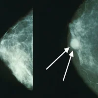
MRT is widely considered as the most
sensitive method to detect breast cancer because, contrary to the conventional
imaging procedures (mammography and ultrasound) it provides functional
information on the blood circulation of the tissue. Generally, this examination
is applied in high-risk patients on an annual basis, who have an increased risk
for breast cancer based on family history or a special mutation (e.g. BRCA 1).
In the process, women are initially
injected with a contrast medium, which renders the so-called “background
parenchymal enhancement” visible in the MRT. It was already possible to
demonstrate in these women that severe background parenchymal enhancement,
which indicates increased blood supply and thus, for example, a hormonally
activated breast tissue, associated with an increased risk of the disease.
Bennani-Baiti and Baltzer, together with
Matthias Dietzel of the University Clinic Erlangen, took up the issue
whether breast tissue with increased background parenchymal enhancement is
generally an indicator for an increased risk for breast cancer, and not just in
the risk group. They conducted a cross-sectional study and analysed the
findings of a group of 540 patients who had been transferred to an MRT for the
further clarification of abnormal findings.
Ultimtely, only the age was decisive for
the risk of breast cancer in this group of patients, who did not have an
increased risk for breast cancer due to familial accumulation or known
mutations. The study data further showed that very active breast tissue in
non-high-risk patients points to a lesser age and thus to a lesser risk for the
illness. It is also not necessary for these women to undergo further
examinations.
However, with their work, Bennani-Baiti,
Dietzel and Baltzer demonstrated the importance of the knowledge of background
parenchymal enhancement for high-risk patients for a risk assessment of breast
cancer. Their results indicate that the changed breast tissue of high-risk
patients is more susceptible to degeneration compared to women without these
risk factors.